
Dose Calibrators & Accessories
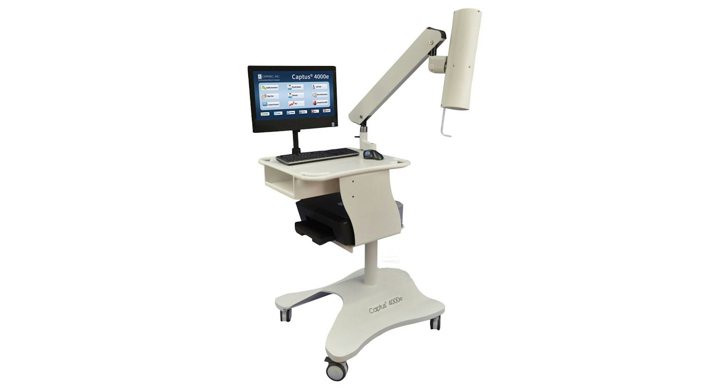
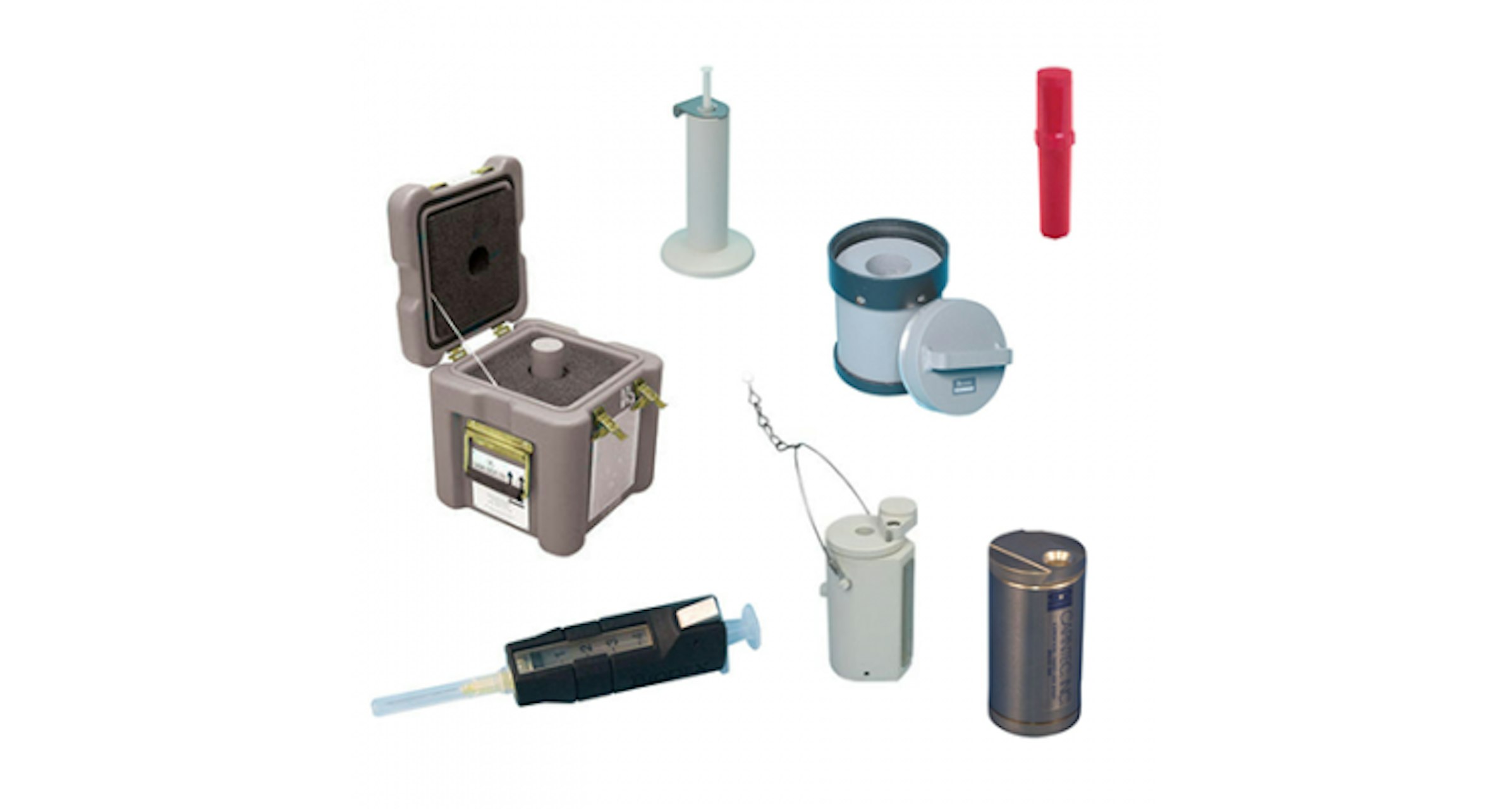
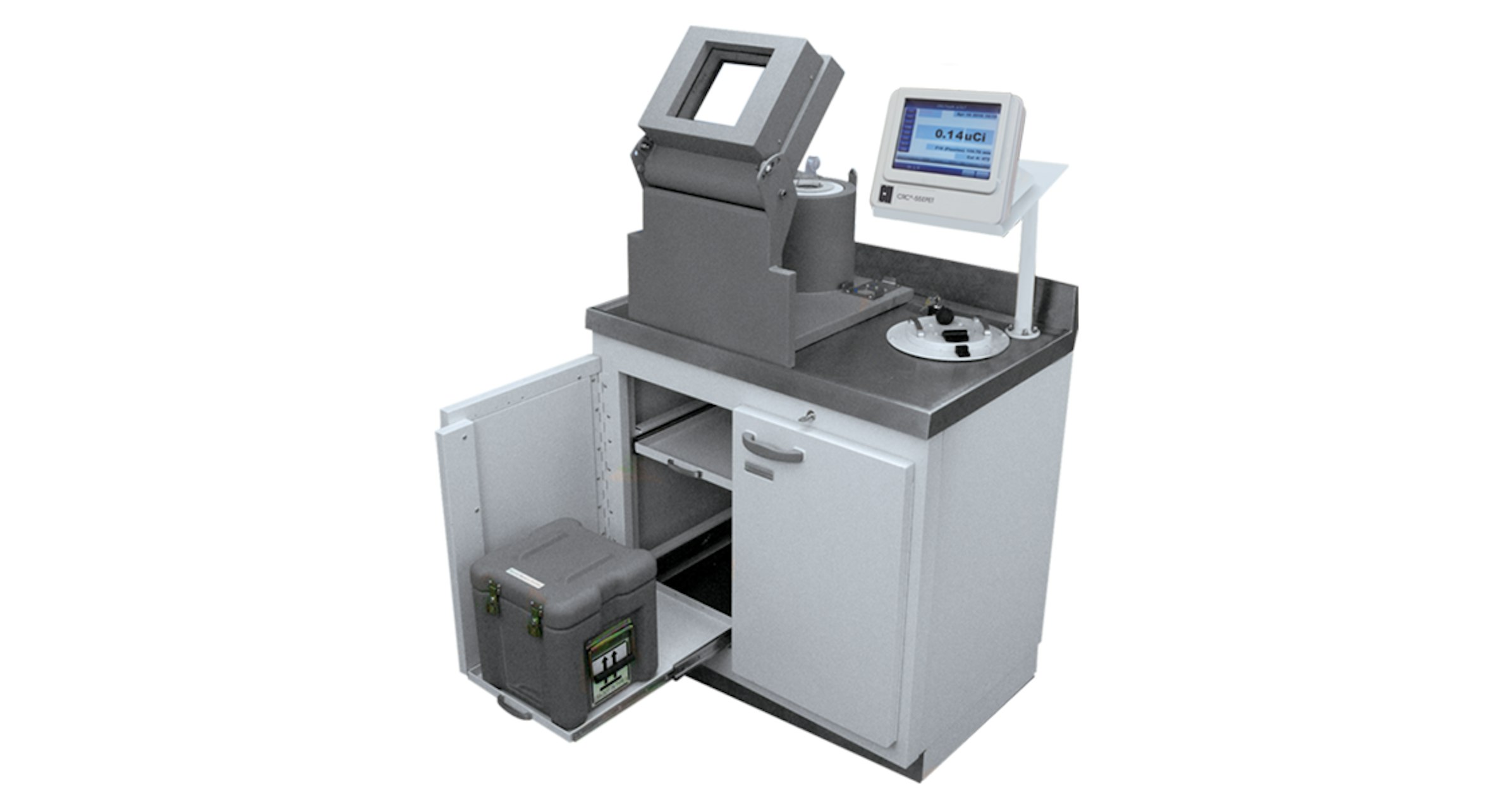
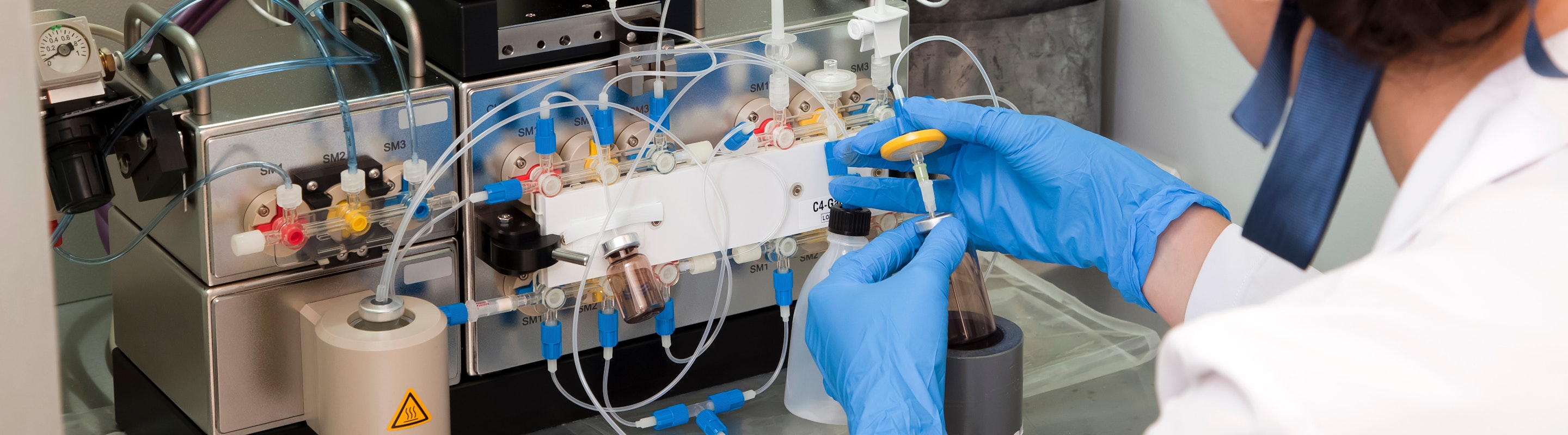
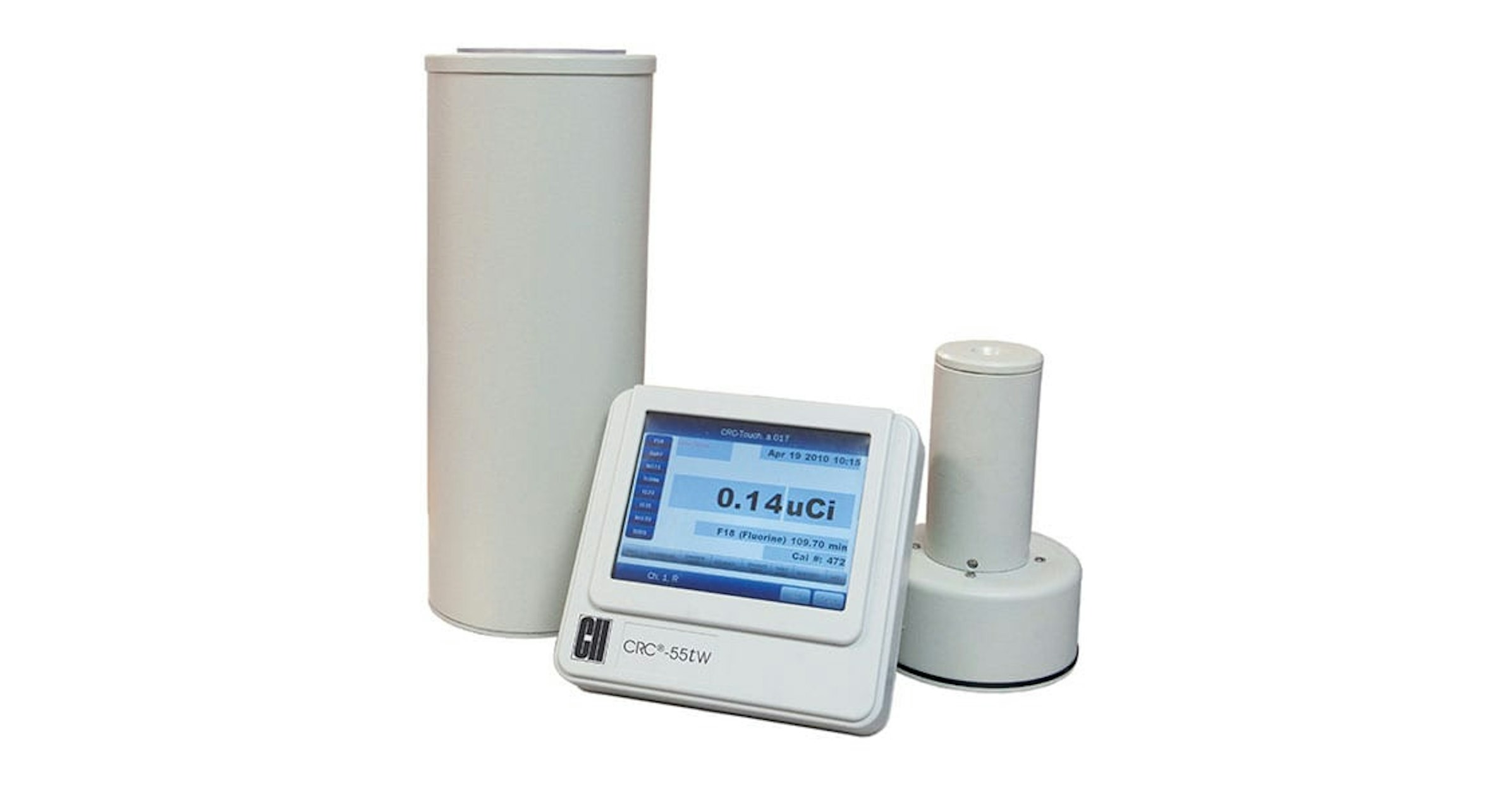
Preparing and administering doses in nuclear medicine require exact measurements, reliability and safe handling. Our Dose Calibrators and Accessories allow for fast, accurate dose preparation, QC and measurement with minimal handling for optimal safety.
Preparing and administering doses in nuclear medicine require exact measurements, reliability and safe handling. Our Dose Calibrators and Accessories allow for fast, accurate dose preparation, QC and measurement with minimal handling for optimal safety.