Bar Phantoms
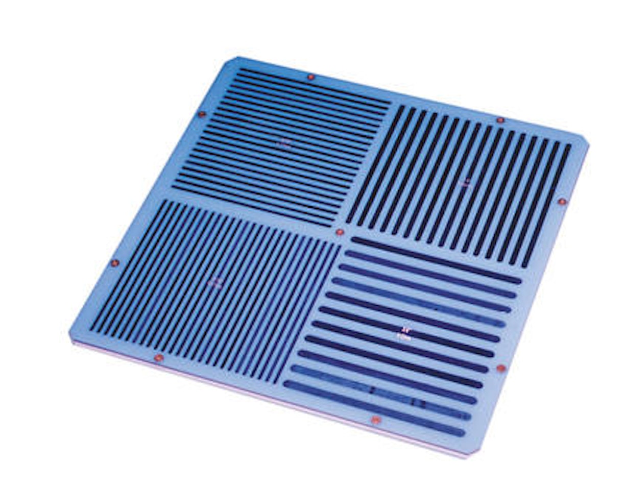
Four-quadrant Bar Phantoms
Four-quadrant Bar Phantoms

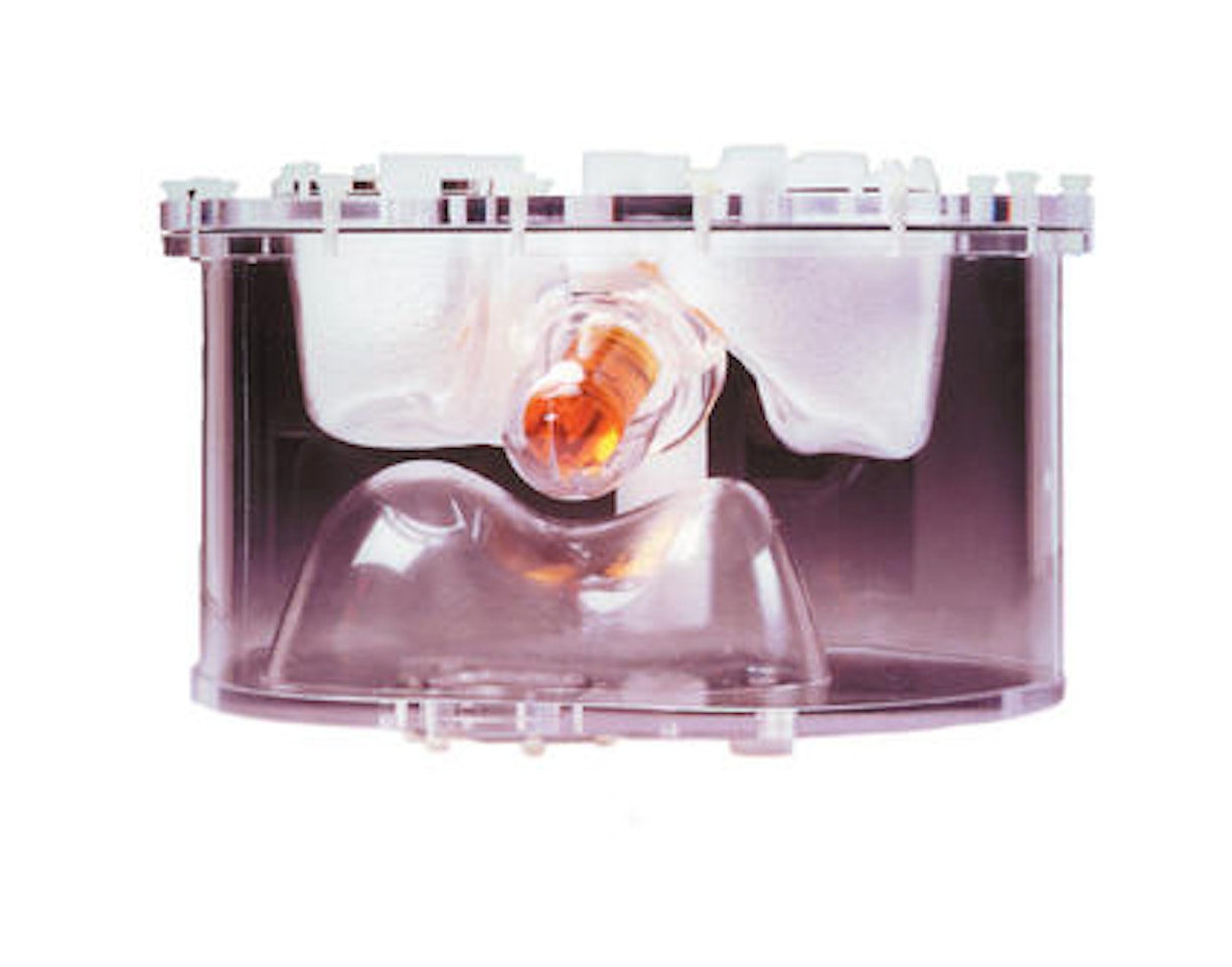
Provides an anatomically accurate three-dimensional simulation of the radioisotope distribution found in the normal brain.
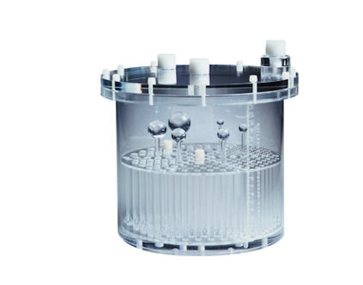
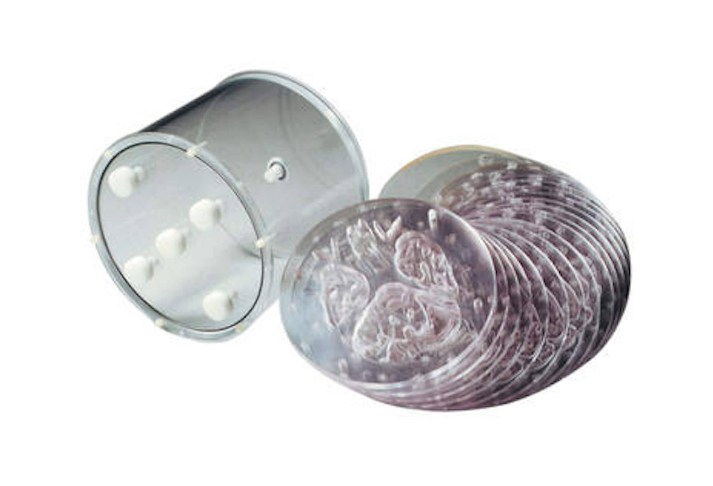
The Hoffman 3-D Brain Phantom provides an anatomically accurate three-dimensional simulation of the radioisotope distribution found in the normal brain. The phantom allows quantitative and qualitative study of the three-dimensional effects of scatter attenuation as they would appear in Iodine-123-IMP or Iodine-123-HIPDM imaging with single photon emission computer tomography or fluorine-FDG-F18 imaging with positron emission computed tomography. The phantom simulates the 4:1 uptake ratio in the gray and white matter, normal in these studies. Ventricles that are normally void of radioactivity are present. The phantom is composed of sturdy plastic and a single fillable chamber that eliminates the necessity of preparing different concentrations of radioisotope. Nineteen independent plates stack neatly within the cylindrical phantom for easy disassembly and assembly. The user can easily add their own custom defects to simulate clinical abnormalities.
The phantom can be filled with the appropriate radioactive material or contrast material for SPECT, PET or MRI applications.
Each insert is made up of five thinner slices. Two slices 0.03″ thick interspersed in 0.6″ thick slices to create a composite slice.
| 043-790 | Phantom, Hoffman 3-D Brain |
Do you have a question or need a custom solution? We're here to help guide your research.
Looking for Services or Support?
We're here to help.